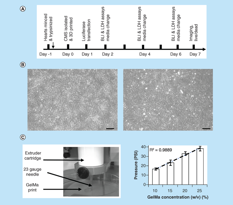
Figure 1. Outline of experimental design.
(A) CMCs and CFBs were isolated using standard procedure that involves overnight digestion with trypsin which followed by collagenase digestion next day. Cells were then mixed with GelMA, printed and placed in culture conditions for specified number of days. On days 2, 4, and 6, LDH and BLI measurements were taken, followed by imaging of live and fixed cells on day 7. (B) Sample phase-contrast images of CMCs (left) and CFBs (right) cultured in monolayers as parallel controls. Scale bar-100 micron. (C) Commercial 3D bioprinter (Allevi) was used to print cell-laden bioinks using specified values of extruder pressure and syringe temperatures. Graph on the right shows minimum pressures required to extrude different GelMA concentrations using 23-gauge needle at 20°C temperature. R stands for Pearson correlation coefficient.
BLI: Bioluminescence intensity; CFB: Cardiac fibroblast; CMC: Cardiac myocyte; GelMA: Gelatin methacryloyl; LDH: Lactate dehydrogenase.

