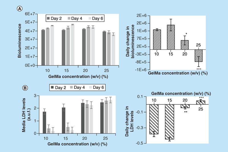
Figure 3. Effects of different gelatin methacryloyl concentration on viability of cardiac fibroblast-laden constructs.
(A) Left panel: values of BLI from 3D printed constructs containing CFBs transfected with luciferin luciferase. The BLI values are expressed in ph/s/cm2/sr or photons per second per centimeter squared per steradian. Right panel: data shown in left panel recalculated as a daily change in BLI. Positive slopes for the 10 and 15% GelMA constructs point to progressive increase in BLI, while constructs printed with 25% GelMA show decline in BLI during 1 week in culture. Asterisks stand for difference with 10% GelMA constructs; *p < 0.05, **p < 0.01, ***p < 0.001. (B) Left panel: values of LDH release into the media from the same 3D printed constructs (arbitrary fluorescence units). Right panel: daily change in LDH release expressed as a slope between LDH and number of days in culture. Negative slopes for the 10 and 15% GelMA constructs point to increasing viability of the cells, while constructs printed using 20 and 25% GelMA show near zero or positive slopes. The latter points to a continuous loss of cells.
BLI: Bioluminescence intensity; CFB: Cardiac fibroblast; GelMA: Gelatin methacryloyl; LDH: Lactate dehydrogenase.

