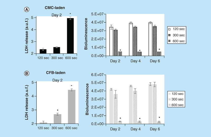
Figure 5. Effects of UV exposure on viability of cardiac myocyte- and cardiac fibroblast-laden, 3D printed constructs.
Immediately after printing, GelMA constructs were exposed to 405 nm, 7 mW/cm2 Watt LED light for either 120, 300 or 600 s. Bioluminescence intensity values from cell-laden 3D printed CMC- and CFB-laden constructs taken at 2, 4 and 6 days of culturing. Initial UV exposures are indicated on x-axis. Shown are the BLI intensities and values of LDH release after 2 days of specified durations of UV exposure for CMC- (A) and CFB-laden constructs (B). Asterisks stand for difference with constructs exposed to UV for 120 s; *p < 0.001.
CFB: Cardiac fibroblast; CMC: Cardiac myocyte; LDH: Lactate dehydrogenase.

