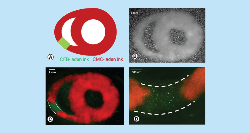
Figure 7. Dual extruder printing using cardiac myocyte- and cardiac fibroblast-laden gelatin methacryloyl.
(A) A cartoon of heart cross-section with scar-like segment which was then designed and assembled in SolidWorks. (B) Visual appearance of a freshly printed construct based on the above schematics using dual extruder printing with CMC- and CFB-laden 15% GelMA bioink formulations. (C) Multispectral imaging of the same construct using 360 nm LED source enabled clear demarcation of the two bioinks (CMCs were labeled with MitoTracker Red, while CFB with Calcein AM). Linear unmixing was applied as per manufacturer software (PerkinElmer Nuance FX 3.02). Dotted white lines outline the CFB-laden part of the construct. (D) A close-up image of CFB segment obtained using Olympus fluorescence microscope. Dotted white lines outline the CFB-laden part of the construct.
CFB: Cardiac fibroblast; CMC: Cardiac myocyte; GelMA: Gelatin methacryloyl.

