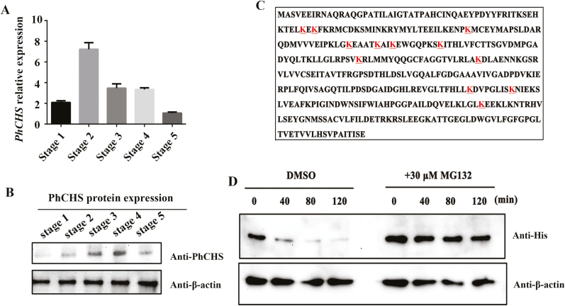
Fig. 6.
PhCHS accumulates in petals and its degradation is mediated by the 26S proteasome system. (A) PhCHS expression from stage 1 to stage 5 of petals (mean ±SD; n=3). (B) PhCHS protein accumulation from stage 1 to stage 5 of petals by western blot analysis; anti-β-actin was used as control. (C) The ubiquitination sites (underlined letters) in PhCHS identified by mass spectroscopy. (D) Cell-free degradation assay of recombinant His–PhCHS protein. Recombinant His–PhCHS was purified from Escherichia coli incubated with petal crude proteins at different stages and treated with specific 26S proteasome inhibitor MG132 at various time intervals. Western blot analysis was conducted using an anti-His antibody, and anti-β-actin protein concentration was used as a loading control. (This figure is available in color at JXB online.)

