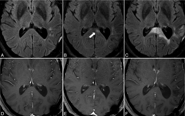
Fig. 5.
Progressive disease suggested by changes on FLAIR images. Serial MR imaging after left parietal lobe GBM resection for a patient treated with anti-VEGF therapy and temozolomide chemotherapy. Small, high-intensity spots are seen on FLAIR imaging (A) at cycle 2; a new high-intensity area (arrow) is found in the left splenium of the corpus callosum on FLAIR imaging (B) at cycle 4 and significantly worsens on FLAIR imaging (C) at cycle 6; No enhancement is detected on Gd-enhanced T1-weighted images (D–F).

