Figure 1.
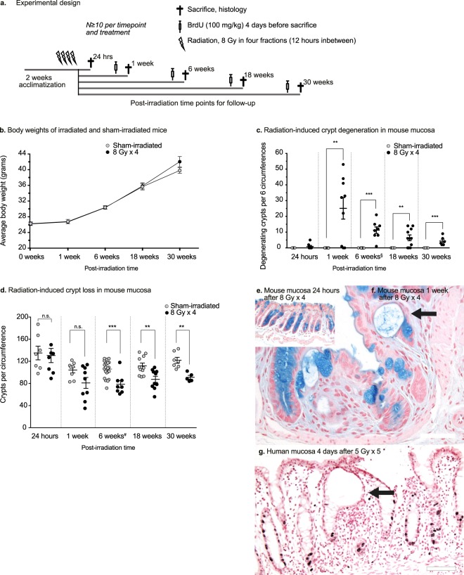
(a) Experimental design. Male C57BL/6J mice (N ≧ 10 per treatment and time point) were irradiated with four fractions of 8 Gy (8 Gy × 4) with 12 hours between each delivered fraction. Four days before sacrifice, the mice were injected with BrdU (100 mg/kg), to follow crypt cell survival. Colorectal tissues were harvested at 24 hours, 1 week, 6 weeks, 18 weeks, and 30 weeks post-irradiation. (b) Weight curves (grams) for the 30-week group. The mice survived and gained weight in a manner similar to the sham-irradiated controls. At 30 weeks post-irradiation, there is a trend towards increased weight in the irradiated mice. (c) Number of degenerating crypts per six circumferences. Radiation-induced crypt degeneration is evident first at 1 week post-irradiation. The number of degenerating crypts diminishes over time. (d) Crypt loss over time. Quantification of the surviving crypts per circumference of the colorectum reveals that the crypt loss is permanent. At 24 hours post-irradiation, no crypt loss is detected. One week after irradiation, a few animals have lost more than 50% of their crypts per circumference, as compared to the sham-irradiated controls, while some animals still have the normal number of crypts. At 6 weeks post-irradiation, crypt loss is seen in nearly all the animals in the irradiated group. The irradiated mucosa has approximately 25% fewer crypts than the sham-irradiated mucosa, and this remains unchanged throughout the experiment, despite what appears to be a slight increase in the number of crypts with age in both groups. §,#Data from the 6-week group have been reported previously26, and9, respectively. (e,f) Histology of irradiated mucosa samples. There are negligible numbers of degenerating crypts 24 hours after irradiation (e). However, at 1 week post-irradiation, several animals have multiple degenerating crypts (f, arrow) with few goblet cells (Alcian Blue and Nuclear Fast Red stain). Scale bars, 50 μm. (g) Degenerating crypt in the rectal mucosa of a cancer patient 4 days after 5 Gy × 5 (G, arrow). There are multiple dividing cells in nearby crypts, visualised as darkly stained Ki-67+ nuclei. Only two Ki-67+ cells are visible at the bottom of the degenerating crypt. Scale bar, 100 μm.

