Figure 3.
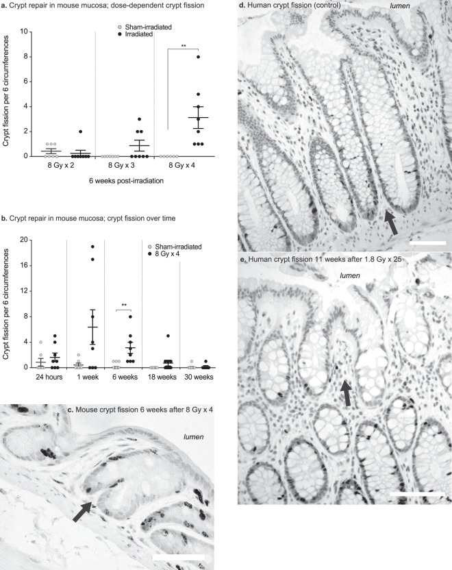
(a) Numbers of crypt fission events per 6 circumferences after different fractionation schedules. The occurrence of crypt fission is dose-dependent, as seen 6 weeks after irradiation. (b) Number of crypt fission events per six circumferences at 24 hours, and at 1 week, 6 weeks, 18 weeks, and 30 weeks after irradiation or sham-irradiation. A few of the irradiated animals show many crypt fission events 1 week after irradiation, and at 6 weeks post-irradiation all the animals exhibit crypt fission; thereafter the number of fission events declines. At 30 weeks post-irradiation, there is no difference in the number of crypt fissions between the irradiated and sham-irradiated animals. (c) Irradiated mouse colorectal mucosa 6 weeks after 8 Gy × 4 of irradiation. The dark nuclei are cells that are labelled with BrdU, showing newly born cells migrating to form two new crypt walls at the crypt mid-line (arrow). (d) Crypt fission in a non-irradiated human rectal biopsy (arrow), showing multiple darkly stained Ki-67+ proliferating cells at the bottom of the dividing crypt. (e) Crypt fission in a human rectal biopsy (arrow) harvested 11 weeks after 1.8 Gy × 25. A few Ki-67+ proliferating cells are seen at the bottom of one of the two resulting crypts.

