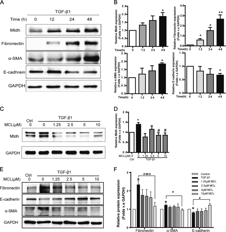
Fig. 5.
DMAMCL/MCL Inhibited both Mtdh and EMT in vitro. a Representative Western blot shows Mtdh and EMT markers (fibronectin, α-SMA, and E-cadherin) in mTECs cell lysates treated with TGF-β1 (5 ng/ml) for 0, 12, 24, 48 h. b Relative levels of the Mtdh and EMT marker proteins compared to GAPDH. *P < 0.05 compared with 0 h; **P < 0.01 compared with 0 h. c mTECs were coincubated with MCL (0, 1.25, 2.5, 5, or 10 μM) and TGF-β1 (5 ng/ml). Cells were harvested 48 h later, and cell lysates were immunoblotted to detect Mtdh expression. d Relative levels of the Mtdh, proteins compared to GAPDH. *P < 0.05 compared with the control group; #P < 0.05 compared with TGF-β1 group. e Representative Western blot on EMT markers in MCL- TGF-β1 coincubation cell lysates samples. f Relative levels of the EMT markers in the blot shown in (e). *P < 0.05 compared with the control group; #P < 0.05 compared with the TGF-β1 group; ####P < 0.0001 compared with the TGF-β1 group. The data are presented as the mean ± SEM of at least three independent experiments

