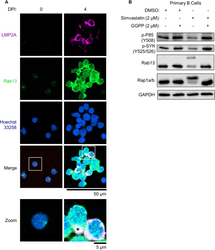
Fig 6. Rab13 colocalizes with LMP2A in newly infected primary B-cells.
(A) Immunofluorescence micrographs of LMP2A, Rab13 and nuclear Hoechst 33528 uninfected versus infected primary human B-cells at 4 DPI. Merged images are shown with white boxes indicating inset images that have been further magnified. The asterisk denotes an area of strong Rab13 and LMP2A colocalization. Scale bars are indicated (50 μm for single-channel and merged images; 5 μm for inset images). See also S7A Fig. (B) Immunoblot analysis of WCL from newly infected primary B-cells cultured in the presence of DMSO, simvastatin (2 μM) or GGPP (2 μM) as indicated from days 2–7 post infection. Shown are representative blots from n = 3 experiments. P85 phospho-tyrosine 508 (Y508) and SYK phospho-tyrosines 525/526 (Y525/526) are markers of their activation, whereas unprenylated Rab13 and Rap1a/b migrate at higher electrophoretic mobilities. See also S8 and S9 Figs.

