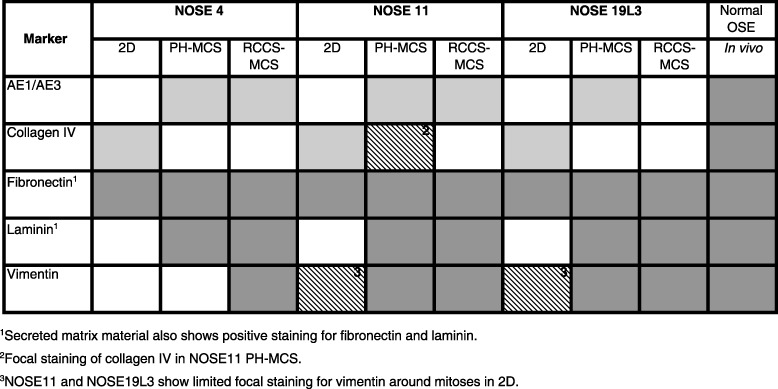
Table 2.
Differential expression of pan‐cytokeratin (AE1/AE3), laminin, vimentin and collagen IV, detected by immunohistochemistry in two‐ (2D) and three‐dimensional (3D) cultures and in primary normal ovarian tissues. Graded shading denotes extent of staining. White denotes negative staining; crosshatched grey denotes weak or focal staining; light grey represents that 20–50% cells stain positive; and dark grey shading indicates over 50% cells stain positive.

