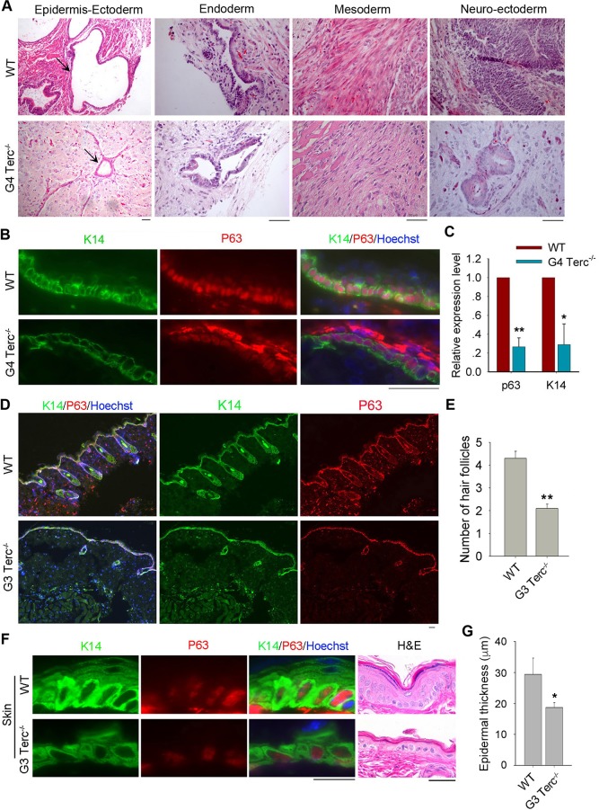
Fig 2. Short telomeres impair epidermal differentiation in vivo.
(A) Three embryonic germ layers shown by histology following H&E staining of teratomas formed from WT and G4 Terc–/–ES cells. Scale bar = 50 μm. (B) Immunofluorescence of epidermis markers shown by K14 and nuclear P63 in teratomas formed from WT and G4 Terc–/–ES cells. Nuclei are stained in blue with Hoechst. Scale bar = 50 μm. (C) Expression levels by qPCR of basal layer markers K14 and p63 in teratomas formed from WT and G4 Terc–/–ES cells. Bars = Mean ± SEM (n = 3). *, p<0.05; **, p<0.01, compared with WT teratomas. (D) Representative immunofluorescence images showing co-staining of P63 with K14 in the sections of mouse skin epidermis. WT mouse skin displays many hair follicles underneath and G3 Terc–/–mouse skin shows fewer and smaller hair follicles. Scale bar = 25 μm. (E) Number of hair follicles in WT and G3 Terc–/–mouse skin per field view. Ten field view was counted, **, p<0.01. (F) Representative images showing skin (back) of WT and G3 Terc–/–mice revealed by immunofluorescence of K14 and P63 and histology by H&E staining. Scale bar = 20 μm. (G) Thickness of skin epidermis in WT and G3 Terc–/–mice estimated from H&E histology. *, p<0.05.

