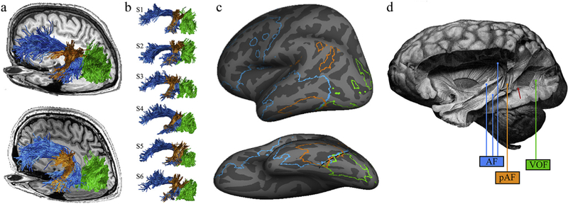
Fig. 1 –

The arcuate fasciculus and the vertical occipital fasciculus. The vertical occipital fasciculus (VOF; green), the arcuate fasciculus (AF; blue) and the posterior segment of the arcuate fasciculus (pAF; orange) are shown in living and post-mortem human brains. (a) The VOF, AF, and pAF are shown for two representative subjects against the background of their T1-weighted anatomy. (b) Renderings of the VOF, AF, and pAF for six additional subjects to illustrate variability. For some subjects, there is a clear separation between the pAF and VOF (S1 and S2), while for others, there is no sharp boundary between the VOF and the pAF (S4 and S5). (c) Cortical endpoints for these three pathways were defined for 37 subjects. Cortical alignment was used to transform each individual’s endpoint map to the FreeSurfer average template (www.freesurfer.net), and regions with consistent, intersubject overlap are shown for each pathway. Outlined cortical extents indicate locations in which the AF, pAF, or VOF terminated in more than 10 subjects. The black dashed line highlights the cortical location where the three pathways converge (posterior occipitotemporal sulcus extending into the lateral fusiform gyrus). (d) Curran’s identification of the VOF, AF, and pAF in the post-mortem human brain (Curran, 1909). Labels have been added for clarity, but Curran distinguished these three tracts. He highlights the fact that the pAF and VOF abut one another, writing: ‘In this dissection, most of this bundle is removed showing a large window through which the fasc. o. f. inf. [IFOF] can be seen. At the anterior of this window a small part of the fasc. trans. occ. [VOF] still remains’. (Bracketed terms have been added to reflect modern terminology). The red arrow highlights the anterior portion of the VOF that Curran describes.
