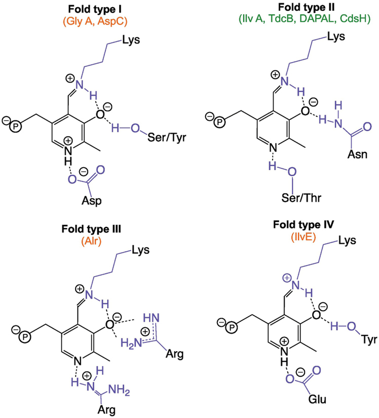
Figure I.
Generalized scheme showing coordination of pyridine N and 3’O from pyridoxal 5’-phosphate (black) for the four main fold types of PLP-dependent enzymes. Since fold types V-VII contain members belonging to only one reaction type, these were excluded from the scheme. Enzyme active site residues are shown in blue and the phosphate group of PLP is shown with ‘℗’. Beneath the fold type label, generators of free 2AA are highlighted in green and enzymes damaged by free 2AA are shown in red. Note, the identified residues are provided using the listed enzymes as templates; for example, fold type III decarboxylases coordinate the pyridine N with a glutamate residue replacing the listed arginine residue. A comprehensive list of PLP coordinating residues is provided by Singh et al. [86].

