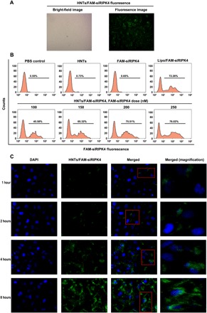
Fig. 2. The transfection efficiency and distribution of HNT-delivered siRIPK4 in T24 bladder cancer cells.

(A) Bright-field and fluorescence microscopy images of HNTs/FAM-siRIPK4 fluorescence at 6 hours after transfection. (B) Flow cytometry analysis of fluorescent cells: representative histograms (upper panels) and means ± SD (lower panels, from three independent experiments). FAM-siRIPK4, fluorescein-labeled siRNA targeting RIPK4. (C) Confocal laser scanning microscopy analysis of the distribution of HNTs/FAM-siRIPK4 in T24 bladder cancer. Fluorescein (green)–labeled siRIPK4. DAPI (4′,6-diamidino-2-phenylindole; blue) was used to stain cell nuclei.
