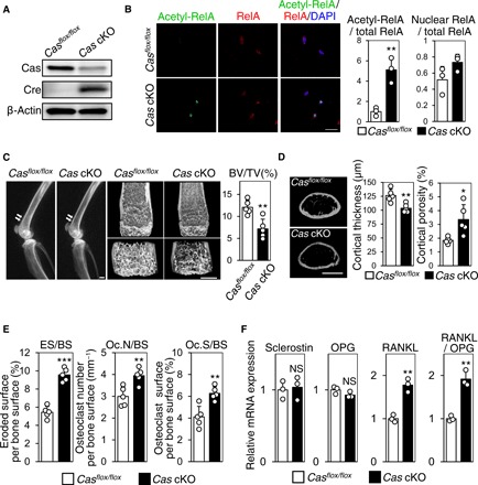
Fig. 2. Increased bone resorption and RANKL expression in osteocyte-specific Cas knockout mice.

(A) Significant reduction of Cas expression in osteocyte fractions derived from Cas cKO mice. (B) Increased RelA acetylation in osteocytes of Cas cKO mice. Confocal images of anti–acetylated RelA (green) and anti-RelA (red) immunofluorescence images of midshaft tibiae of Casflox/flox mice and Cas cKO mice are shown with nuclear staining (left). RelA acetylation (center) and nuclear distribution (right) were quantified as in Fig. 1C. RelA acetylation was scaled with the mean value of control bones (column 1) set at 1 (n = 3 mice per group; each value was averaged from four to six osteocytes analyzed in each bone). Scale bar, 10 μm. (C) Reduced bone mass in Cas cKO mice. Representative radiographic images of the femurs (left; arrows point to apparent differences in bone density) and distal femur μCT (center) of Cas cKO mice and normal Casflox/flox littermates are shown. Scale bars, 1 mm. The ratio of bone volume over total volume (BV/TV) was calculated from μCT images (right, n = 5 mice per group). (D) Loss of cortical bone in Cas cKO mice. Representative μCT images of femoral cortices are shown. Scale bar, 1 mm (left). Cortical thickness (center) and cortical porosity (right) were calculated from μCT images (n = 5 mice per group). (E) Increased osteoclastic bone resorption in Cas cKO mice. Parameters for osteoclastic bone resorption in Cas cKO mice and their normal Casflox/flox littermates were determined by histomorphometric analysis (n = 5 mice per group). (F) Increased RANKL expression in osteocytes of Cas cKO mice. mRNA expression levels of sclerostin, OPG, and RANKL as well as RANKL/OPG ratio in the osteocyte fractions derived from Casflox/flox and Cas cKO mice were normalized against data from Casflox/flox mice, which were set at 1 (n = 3 mice per group).
