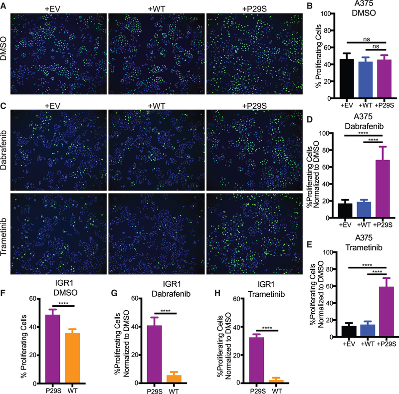
Figure 2. Rac1P29S Confers a Proliferative Advantage to Melanoma Cells upon MAPKi Treatment.

(A-E) Incorporation of fluorescently labeled Edu Into S-phase nuclei of A375 cells exogenously expressing Rac1P29S (+P29S), Rac1WT (+WT), or empty vector (+EV) to detect:
(A and C) proliferating cells (green) and Hoechst-labeled nuclei (blue) to determine fraction of proliferating cells, denoted in %, upon 48 h treatment with (A and B) DMSO (0.003%), (C-E) Dabrafenib (33.3 nM), and Trametinib (3.3 nM).
(F-H) Proliferation assay applied to IGR1 cells endogenously expressing Rac1WT or Rac1P29S upon 48 h treatment with (F) DMSO (0.1%), (G) Dabrafenib (10,000 nM), and (H) Trametinib (6.6 nM).
For all Edu incorporation assays n = 15 images were counted for each condition from three independent experiments. All data represent mean ± SD. Statistical significance was assessed by one-way ANOVA with Tukey’s multiple comparisons test (B, D, and E) and unpaired Student’s t-test (F, G, and H). ****p < 0.0001.
See also Figure S2.
