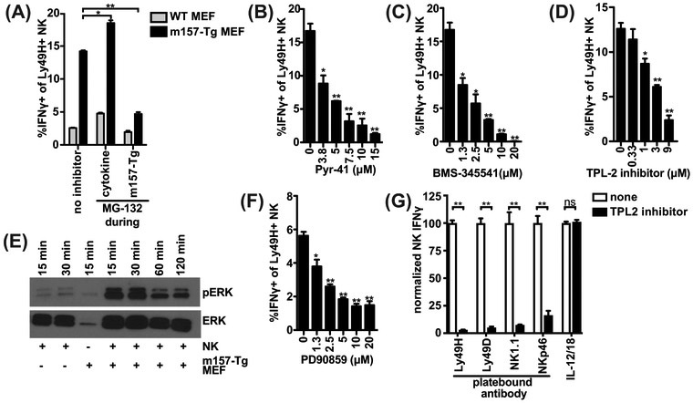
Figure 5: Ly49H signaling requires the Proteasome-Ubiquitin-IKK-TPL2-ERK axis to induce IFNγ.
(A) As in Figure 4AB, splenocytes were pretreated with IL-12, hereafter the cells were washed and incubated with m157-Tg MEF in the presence of monensin. During IL-12 (cytokine) or m157-Tg stimulation, the proteasome inhibitor MG-132 (10μM) was added. (B-D,F) as in Figure 4E, splenocytes were pretreated with IL-12, thereafter the cells were washed and stimulated with m157-Tg MEF, in the presence of monensin and E1 inhibitor Pyr-41 (B), IKK-complex inhibitor BMS-345541(C), TPL2 inhibitor (D), or ERK-inhibitor PD90859 (F) at the indicated concentrations. (E) Purified NK cells were pre-stimulated with IL-12, after which the NK cells were stimulated with m157-Tg MEF for the indicated time. ERK and phospho-ERK levels were analyzed by western blot. (G) Splenocytes were stimulated with indicated plate-bound antibody or IL-12 (12.5 ng/ml) and IL-18 (5 ng/ml) in the presence or absence of 9 μM TPL2 inhibitor. Percent of IFNγ producing NK cells were normalized to the condition without inhibitor. Representative experiments of 2 (A, D, E, G) or 3 (B, C, F) independent experiments are shown and were performed in duplicate (A, B, C, F, G) or triplicate (D). **p<0.01, *p<0.05, ns= not significant.

