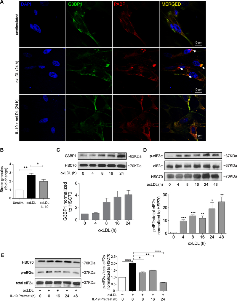
Figure 3.

OxLDL induces stress granule formation in human VSMCs. (A) Representative images of human VSMC either unstimulated or treated with oxLDL (50 μg/mL) and stained for the stress granule marker PABP (red) or alpha actin (green). Cell nuclei were stained with DAPI (blue). Arrows indicate PABP+ puncta. (B) Quantification of punctate immunoreactivity in unstimulated and oxLDL-treated human VSMC. (C) Western blot analysis of G3BP1 and HSC70 protein levels in human VSMCs treated with oxLDL for the indicated times. Densitometry analysis of G3BP1 protein relative to HSC70 is shown below. Data are the mean of 3 independent experiments. (D) Western blot analysis of total and phospho-eIF-2α in unstimulated and oxLDL-treated human VSMC, densitometry of phospho-to-total protein ratios are shown below. (E) Western blot analysis of total and phospho-eIF-2α in VSMCs untreated or pretreated with IL-19 prior to stimulation with oxLDL. Data are representative of 3 independent experiments. *P≤0.05.
