Figure 5.

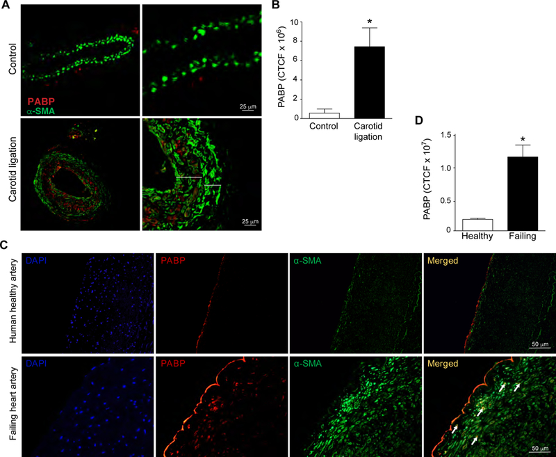
Enhanced PABP expression in injured mouse and human arteries. (A) Representative images of control and ligated mouse carotid arteries stained for PABP (red) and alpha smooth muscle actin (green) Larger bracket indicates neointima, smaller bracket is the media. (B) Quantification of PABP immunoreactivity in control or ligated mouse carotid arteries. N=4 mice/group. (C) Representative images of normal human coronary artery and coronary artery from a failing human heart stained for PABP (red), alpha smooth muscle actin (green), and DAPI (blue). (D) Quantification of PABP immunoreactivity in human artery samples expressed as corrected total cellular fluorescence (CTCF). n=3 control, n=4 failing heart.
