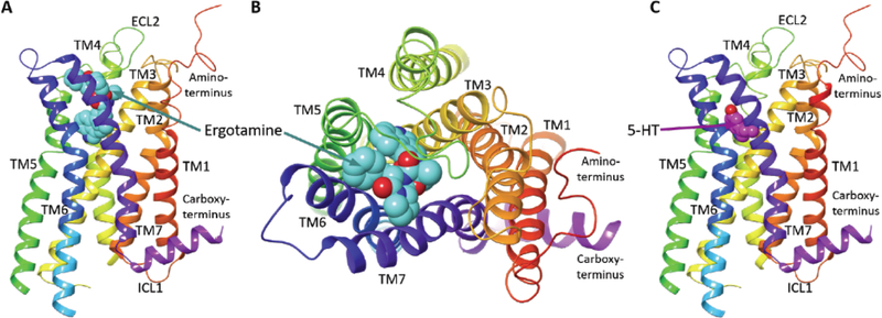
Fig. (1).
The recently solved X-ray crystal structure of the “active-like” state of the 5-HT2CR bound to ergotamine (PDB code: 6BQG) and 5-HT2CR model bound to 5-HT is depicted. (A) The side view of the ergotamine-bound 5-HT2CR crystal structure with the N- and C-termini, extracellular and intracellular loops, and the transmembrane helices labeled; ECL = extracellular loop, ICL = intracellular loop, TM = transmembrane domain helix, ergotamine (cyan space fill representation) at the orthosteric site [5]. (B) Top/extracellular-view of the ergotamine binding site. (C) The side view of a 5-HT2CR structure model based on the crystal structure with 5-HT docked to the orthosteric site; 5-HT (magenta space fill representation). The 5-HT-bound 5-HT2CR was generated via induced fit docking protocols on 6BQG using the Schrödinger Drug Discovery Suite. All ligands shown in space filling representation and CPK coloring scheme for N and O atoms.

