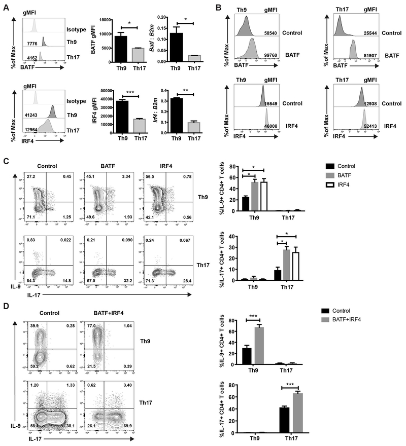
Fig. 1. BATF promotes lineage specific cytokine production in Th9 and Th17 subsets.
(A-C) Naive CD4 T cells were cultured under Th9 or Th17 conditions. Cells were harvested on day 5. (A) BATF and IRF4 expression was detected by intracellular staining and qRT-PCR. RNA expression data was normalized to B2m expression. (B and C) Cells were transduced with MIEG-GFP, BATF-GFP or IRF4-GFP expressing retrovirus on day 1. Transcription factor expression and cytokine expression was detected on day 5 after PMA/ionomycin stimulation. Dot plots were gated on cells that were live CD4+GFP+ cells. (D) Cells were co-transduced with MSCV-Thy1.1 and MIEG-GFP control or BATF-Thy1.1 and IRF4-GFP expressing retrovirus. Cytokine production was detected on day 5 after PMA/ionomycin stimulation. Dot plots were gated on live CD4+Thy1.1+GFP+ cells. Date are mean ± SEM of 3 mice per experiment and representative of three independent experiments. A two tailed Student’s t-test was used for pairwise comparisons. One-way ANOVA with a post-hoc Tukey test was used to generate p-values for all multiple comparisons. *, p<0.05, **p<0.01, ***p<0.001.

