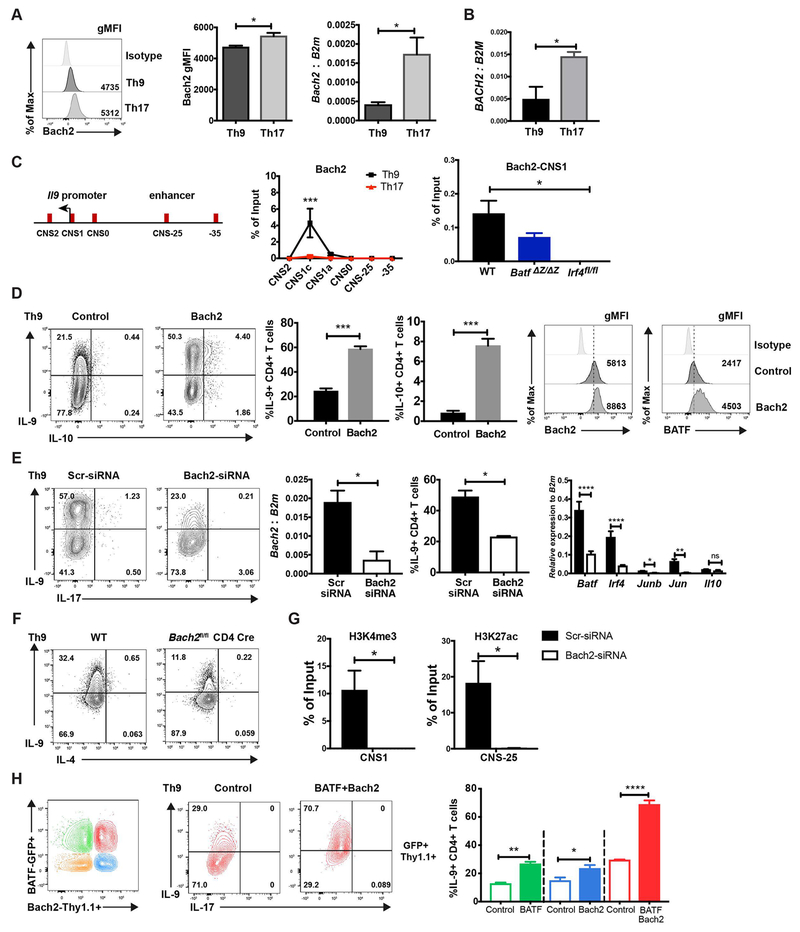
Fig 4. Bach2 promotes IL-9 production in Th9 cells.
(A) Naive mouse CD4 T cells were cultured under Th9 and Th17 conditions. Bach2 expression was detected by ICS (mouse) and qRT-PCR (mouse and human) on day 5. (B) Naïve human CD4 T cells were cultured under Th9 and Th17 conditions and BACH2 expression was detected by qRT-PCR. RNA expression data was normalized to B2M expression. (C) ChIP assay of Bach2 binding to the Il9 gene locus in WT, BATF- and IRF4-deficient Th9 and WT Th17 cells was performed on day 5. (D) MSCV-Thy1.1 or Bach2-Thy1.1 expressing retrovirus was transduced in Th9 cells on day 1. Cells were collected on day 5 for transcription factor staining. Cytokine production was detected after 5 hours PMA/ionomycin stimulation on day 5. Dot plots were gated on live CD4+ Thy1.1+ cells. (E) WT naïve CD4 cells were cultured under Th9 condition with anti-IL-10R and scrambled siRNA or Bach2-specific siRNA was transfected on day 1. Bach2 mRNA expression was analyzed by RT-qPCR after 48 hours transfection. Additional gene expression was performed on day 5 of culture. Cytokine production was detected after 5 hours PMA/ionomycin stimulation on day 4. Dot plots were gated on live CD4+ cells. (F) Naïve CD4 T cells from wild type and Bach2 conditional mutant mice were cultured under Th9 conditions and analyzed for cytokine production as in (E). (G) Cells generated as in (E) were used for ChIP assay of H3K4me3 and H3K27ac at the Il9 locus, performed on day 5. (H) Naive CD4 T cells were cultured under Th9 conditions. MIEG-GFP and MSCV-Thy1.1 (open box) or BATF-GFP and Bach2-Thy1.1 (closed box) expressing retrovirus was co-transduced into the Th9 cells on day1. Cytokine production was detected after 5 hours PMA/ionomycin stimulation on day 5. Dot plots were gated on live CD4+ transduced cells. Data are mean ± SEM of 3 mice per experiment and representative of three independent experiments. *p<0.05, **p<0.01, ***p<0.001, ****p<0.0001.

