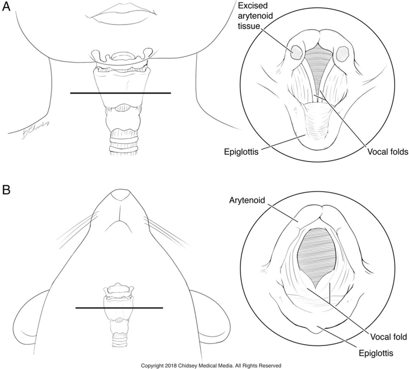
Fig. 1.

Comparison of laryngeal anatomy between human infants and mice. (A) Frontal view showing the relative position and anatomy of the larynx in an infant. The horizontal line indicates the level of the panel at right depicting the location of tissue procurement in human infants undergoing supraglottoplasty. (B) Ventral view of a mouse showing the position of the larynx. Inset at right shows an equivalent view of the mouse larynx indicating the relative position of the arytenoids and other key laryngeal structures. Printed with permission. © 2018 Chidsey Medical Media. All Rights Reserved.
