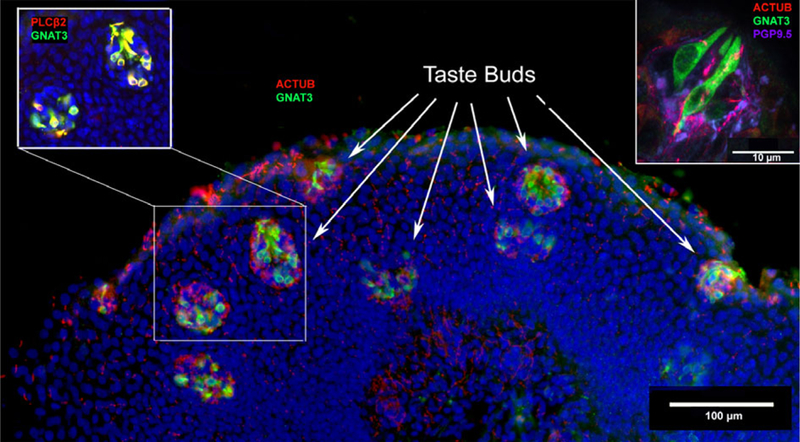
Fig. 2.

Taste buds in human arytenoid tissue. The main figure is a section from a full-term, 5-month-old infant. Staining for markers of type II taste cells, GNAT3 (green) and PLCβ2 (red, upper left inset), show numerous taste buds within a small area of the epithelium. An antibody directed against acetylated tubulin (red, main figure) demonstrates dense innervation of immunoreactive taste cells. In some regions of the epithelium the taste buds are densely packed, making up a density of approximately five taste buds per 100 μm2. Upper right: confocal image taken from the arytenoid tissue of a 34-year-old male donor. Type II taste cells (GNAT3-positive = green) are innervated by nerve fibers shown by acetylated tubulin (red) staining and occur within epithelium that is densely innervated with PGP9.5-positive (magenta) nerve fibers.
