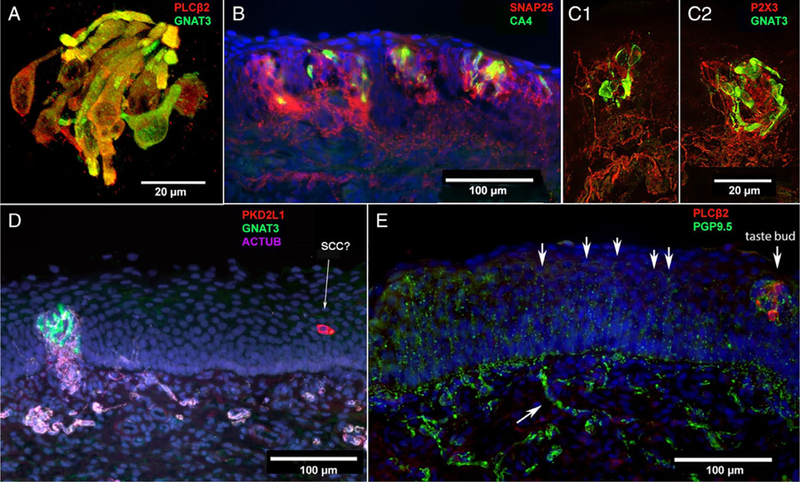
Fig. 3.

Human infant arytenoid taste buds, taste cells, and polymodal nociceptive nerve fibers. (A) Confocal image from a 5-month-old, fullterm infant showing near complete colocalization of GNAT3 (green) and PLCβ2 (red) staining. This suggests that gustducin is the predominant taste-associated G-protein in these tissues. (B) Taste buds from a full-term, 3-month-old infant. Few taste cells express CA4 (green), a marker for sour-responsive type III taste cells. SNAP25 (red) stains nerve fibers showing dense innervation of taste buds. (C1, C2) Two adjacent taste buds from a full-term, 3-month-old infant. Confocal imaging shows dense innervation of GNAT3-positive (green) type II taste cells within taste buds by P2X3-positive (red) nerve fibers. (D) Image from an 8-month-old, premature infant showing a densely innervated (nerve fibers from acetylated tubulin staining = pink) taste bud with GNAT3-positive (green) type II taste cells. Nearby in the epithelium, a PKD2L1-positive (red) solitary cell that is not innervated by acetylated tubulin-positive (magenta) neural fibers. (E) At right is a taste bud with PLCβ2-positive (red) taste cells, closely associated with PGP9.5-positive (green) nerve fibers. At left, numerous free nerve endings, also stained by PGP9.5, extend upward ending near the luminal surface of the epithelium (arrows). The lower arrow indicates a subepithelial axon bundle.
