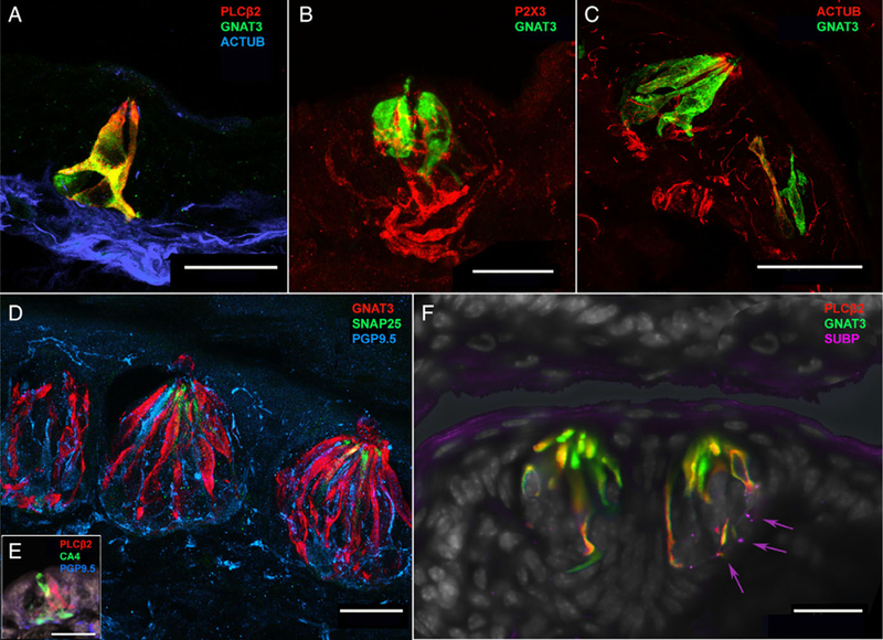
Fig. 4.

Mouse arytenoid taste buds, taste cells, and polymodal nociceptive nerve fibers. (A) Confocal image from a 15-day-old mouse pup depicting a taste bud with near complete colocalization of GNAT3 (green) and PLCβ2 (red), along with innervation by acetylated tubulinpositive (blue) nerve fibers. (B) Confocal image of a taste bud from 15-day-old mouse pup depicting GNAT3-positive (green) type II taste cells innervated by P2X3-positive (red) nerve fibers. (C) Confocal image depicting innervation of a taste bud by acetylated tubulin (red) in a 2-month-old (adult) mouse. Although there are more type II taste cells in the bud (green), the innervation pattern is similar to the 15-day-old mouse shown in (A). (D) Confocal image of three adjacent taste buds situated just proximal to the esophageal face of the arytenoid complex in a 2-month-old (adult) mouse. These taste buds include multiple GNAT3-positive (red) type II taste cells, some SNAP25-positive (green) type III taste cells, and dense innervation by PGP9.5-positive (blue) nerve fibers. PGP9.5-positive nerve fibers also innervate the epithelium surrounding the taste buds reaching near the surface of the tissue. (E) Confocal image from a 15-day-old mouse pup showing CA4-positive (type III) cells adjacent to PLCβ2-positive (type II) cells within a taste bud. (F) Two adjacent taste buds on the esophageal-adjacent surface of the arytenoid in a 2-month-old (adult) mouse. As with the 15-day-old mouse, taste buds shown in (A) and the human infant taste buds in Figure 3A, GNAT3 and PLCβ2 demonstrate near-complete colocalization in type II taste cells. These taste buds are also innervated by small substance P-positive (magenta) neural fibers (arrows).
