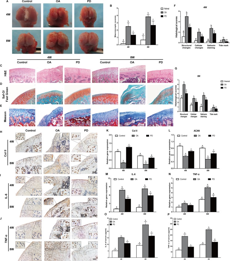
Figure 4.
Protection effects of PD on the treatment of OA. In vivo cartilage repair at 4 and 8 weeks post-surgery, (A) Macroscopic appearance and (B) ICRS scores of femoral condyles from OA rat were detected. (C–E) H&E staining, Saf-O/Fast Green staining, and Masson staining were performed in sections of cartilage. Original magnification × 40 (scale bar, 2,000 μm). (F,G) Histological score of articular cartilage was determined. (H–J) Immunohistochemical staining of Col II, IL-6, and TNF-α was performed in sections of cartilage. Original magnification × 80 and × 320. (K–N) qRT-PCR was performed to analyze the expression of Col II, ACAN, TNF-α, and IL-6 genes in cartilage at 4 and 8 weeks. Meanwhile, serum levels of (O) IL-6, and (P) TNF-α were determined using ELISA kits. Values are the means ± SD (n = 10). Bars with different letters are significantly different from each other at p < 0.05 and those with the same letter exhibit no significant difference.

