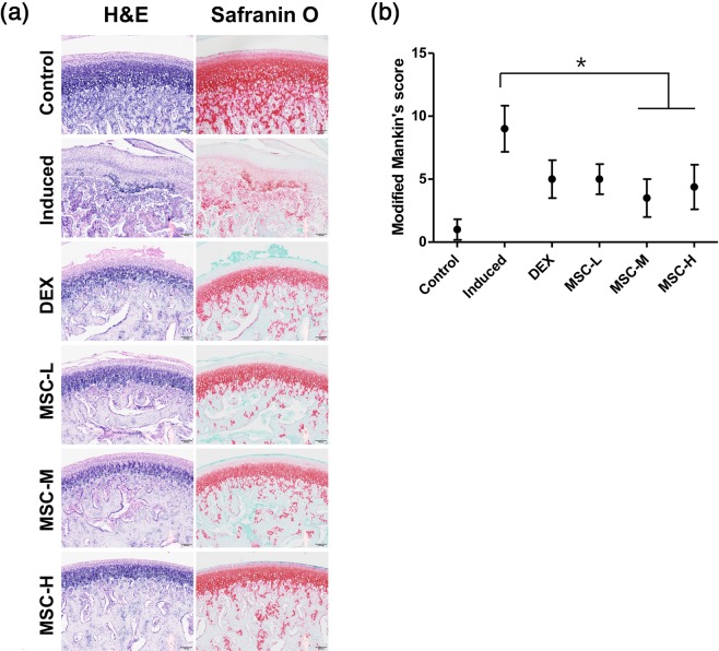
Figure 4.
Cartilage regeneration following transplantation of MSCs derived from human umbilical cord. (a) Histological images showing fewer surface changes and reduced loss of proteoglycans in the MSC-treated groups compared with those in the untreated TMJ-OA-induced group. The untreated TMJ-OA-induced group demonstrated loss of chondrocytes, cellular disarrangement, abnormal thickening of the fibrous layer, changes in cellularity, disruption of osteochondral junction, necrotic bone debris surrounded by fibrous tissue and osteoclasts, and severe loss of SO staining. The DEX-treated group showed fibrillation of the cartilage surface, whereas MSC-treated groups showed a clear surface and mildly reduced SO staining. Scale bar = 100 μm. (b) Modified Mankin scores were significantly higher in the untreated TMJ-OA-induced group than in the control group, but lower in the MSC-treated groups than in the untreated TMJ-OA-induced group. No differences in Mankin scores were noted between the MSC-treated groups. Biological replicates, n = 5; technical replicates, n = 2. Data are represented as mean ± SD. *p < 0.05. MSCs: mesenchymal stem cells; TMJ-OA: temporomandibular joint osteoarthritis; SO: Safranin O, DEX: dexamethasone.

