Figure 2.

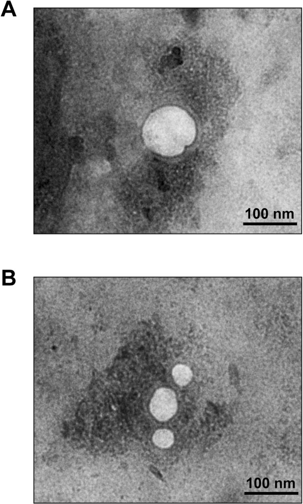
Transmission electron microscopy (TEM). After isolation/purification, urinary microvesicles (A) and exosomes (B) were negatively stained by uranyl acetate and their images were captured by TEM (original magnification = 50,000×).

Transmission electron microscopy (TEM). After isolation/purification, urinary microvesicles (A) and exosomes (B) were negatively stained by uranyl acetate and their images were captured by TEM (original magnification = 50,000×).