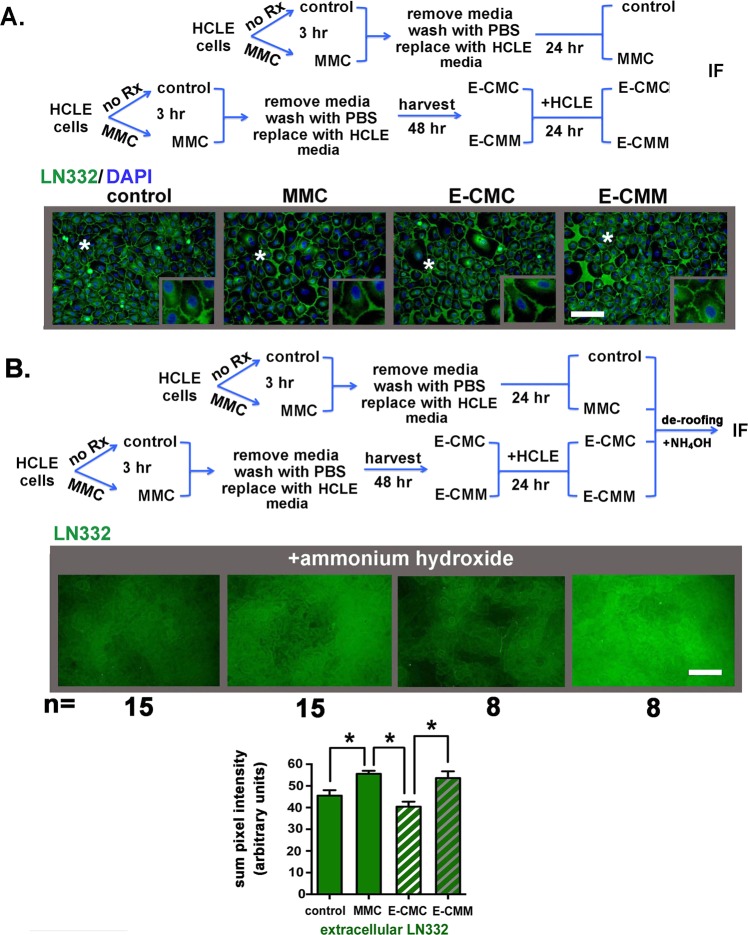
Figure 3.
Transient MMC treatment and growing cells in E-CMM increases the extracellular deposition of LN332 by HCLE cells. (A) Indirect immunofluorescence was performed to assess the localization and expression of the α3β1 and α6β4 integrin ligand LΝ332 in HCLE cells treated as described in the schematic. LN332 appears excluded from the basal surface of HCLE cells. (B) To determine whether the absence of LN332 underneath HCLE cells is due to tight adhesion of cells to LN332 and the inability of the LN332 antibody to reach its epitope, cells grown as described in the schematic were treated with ammonium hydroxide to remove cells, a method referred to as de-roofing, which leaves the ECM behind. Indirect immunofluorescence was performed to assess the deposition of LΝ332 by HCLE cells treated with MMC for 3 hours and in HCLE cells incubated in E-CMC and E-CMM. Deposition of LN332 by HCLE cells was quantified in 15 separate fields in 3 separate wells for control and MMC treated cells and in 8 separate fields in 3 separate wells for E-CMC and E-CMM treated cells. Data are expressed as the summation of pixel intensities per field. Data show that LN332 is excluded from the basal surface of the epithelial basal cells and there is more LN332 deposited by MMC and E-CMM treated cells compared to control and E-CMC treated cells. The magnification bar equals 40 μm.

