Figure 6.
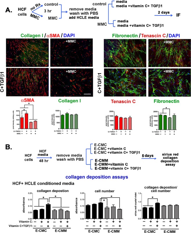
MMC reduces αSMA and fibronectin localization and expression in HCFs and conditioned media from MMC treated HCLE cells blocks TGFβ1-induced collagen deposition. (A) As indicated in the schematic, control and MMC-treated HCFs were grown in HCF media and in media supplemented with vitamin C and TGFβ1 for 48 hours. Cells were fixed, permeabilized and stained with antibodies against αSMA (red) and collagen I (green) and TN-C (red) and fibronectin (green). In addition, nuclei were stained with DAPI (blue). Data show that 48 hours after MMC treatment, αSMA expression and FN fibril formation in HCFs is reduced. The magnification bar = 40 μm. (B) To determine whether exposure to proteins secreted by MMC-treated HCLE cells impacts collagen deposition by HCFs, E-CMC and E-CMM was added to HCF cultures and collagen deposition assessed. Data show that the addition of vitamin C and TGFβ1 to E-CMM did not induce collagen deposition but addition of vitamin C and TGFβ1 to E-CMC increased collagen deposition by HCFs significantly.

