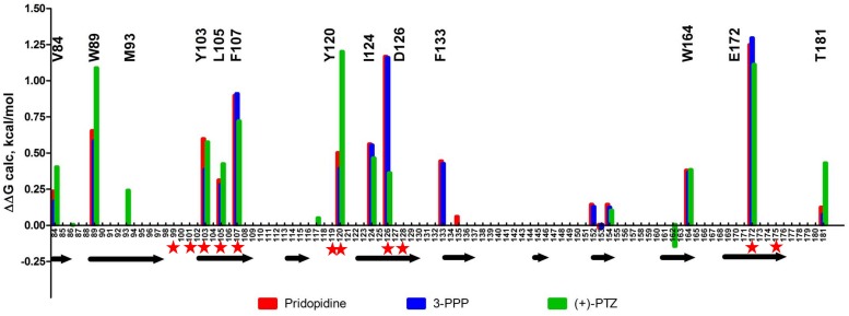
FIGURE 3.
In silico alanine mutagenesis of S1R structures bound to pridopidine, (+)-3-PPP, and (+)-PTZ ligands. Bars indicate energetic contributions of individual residues involved in the formation of ligand binding pocket (red for pridopidine, blue for 3-PPP, and green for PTZ). Critical residues are marked on top of the graph. Secondary structure assignment is given below with each arrow corresponding to a beta-barrel forming strand. Previously reported critical ligand-binding residues are marked with red asterisks.

