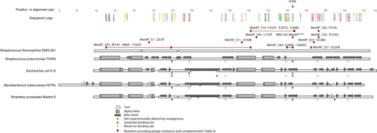
Figure 1.
Protein alignment of S. thermophilus SMQ-301 MetAP with 4 other MetAP for which the structure is available. The WebLogo was only kept when the residue was conserved in all sequences. The secondary structures are represented over each sequence and diamonds indicate active sites and substrate binding sites. Red boxes highlight the mutations that provide phage resistance listed in Table 4. When linked, they occurred in the same MetAP mutant of S. thermophilus SMQ-301. Uniprot accession number of the protein sequences: E. coli K12 (P0AE18), Rickettsia prowazekii Madrid E (Q9ZCD3), S. pneumonia TIGR4 (B2IQ22) and M. tuberculosis H37Rv (P9WK19).

