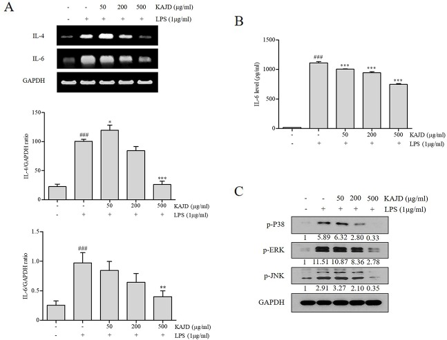
Figure 7.
KAJD inhibits MAPK pathways and suppresses the mRNA and protein expression of proinflammatory molecules in splenocytes. (A) The IL-4 and IL-6 mRNA levels in splenocytes were measured by RT-PCR analysis. The bar graphs represent the quantitation of RT-PCR data. (B) The level of IL-6 in cell culture supernatants was measured by ELISA. Splenocytes stimulated with LPS for 1 h were treated with varying concentrations of KAJD for 24 h. (C) Phosphorylated ERK1/2, p38, and JNK levels in cell lysates were determined by immunoblot analysis. GAPDH was used as an internal control. Data are presented as the mean ± SEM. ###P < 0.001 compared to nonstimulated cells. *P < 0.05, **P < 0.01, and ***P < 0.001 compared to stimulated cells. LPS, lipopolysaccharide.

