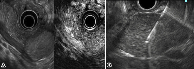
Figure 3.
EUS revealed a well-defined hypoechoic mass region in the pancreatic tail, and contrast-enhanced harmonic EUS using a Sonazoid® showed the tumor as iso-enhanced compared to the surrounding pancreatic tissue (A). The mass was relatively hard; however, the puncture was achieved with little effort, and the needle never bent during the procedures (B).

