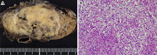
Figure 6.
The surgical specimen showed a well-circumscribed, yellowish-white mass that measured 43mm×30mm and was surrounded by a complete fibrous capsule with a negative surgical margin (A). At the microscopic level, the tumor was composed of spindle-shaped cells possessing clear to focally granular eosinophilic cytoplasm without necrosis, atypia, or frequent mitoses (B) (Hematoxylin and Eosin staining ×200).

