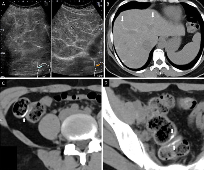
Figure 1.
The abdominal ultrasonography (US) and computed tomography (CT) findings. (A) US (B-mode) revealed a network calcification pattern (white arrows). (B) Non-contrast computed tomography showed calcification along the liver vessel. (C) CT showed calcification in the ascending colon. (D) CT showed calcification in the rectal wall.

