Figure 1.

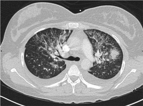
Computed axial tomography of the chest performed with intravenous contrast showing bilateral nodular opacifications in a bronchovascular distribution.

Computed axial tomography of the chest performed with intravenous contrast showing bilateral nodular opacifications in a bronchovascular distribution.