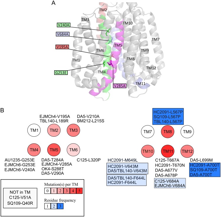
FIG 2.
(A) Mapping of the mutations found in spontaneous EJMCh4- and EJMCh6-resistant mutants on an M. tuberculosis MmpL3 three-dimensional homology model. Only the transmembrane helices (TM) are depicted. The residues mutated in the spontaneous resistant mutants are shown as sticks, and the mutations are indicated in the colored boxes. All TM domains are in gray, with the exception of those carrying a mutation in resistant mutants, notably TM2 (red), TM4 (green), TM5 (magenta), and TM11 (blue). (B) All mutations found in MmpL3 from resistant mutants selected on several MmpL3 inhibitors. Transmembrane helices are represented by circles. Each mutation as well as which drug it confers resistance to is indicated. The number of mutations affecting TM is shown from white (no mutation) to dark red (five mutations), and the blue color indicates residues that confer cross-resistance to several drugs.

