Figure 4.
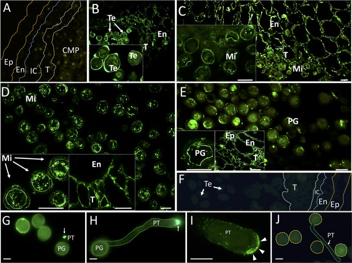
Fluorescence microscopy localization of OeRbohH in the olive anther and in vitro germinated pollen grains. Sections from olive anthers at the following stages: pollen mother cells prior to meiosis (A), tetrads (B), young microspores (C), vacuolated microspores (D), and mature pollen (E) were incubated with an anti-OeRbohH Ab, followed by an anti-chicken IgG-Alexa Fluor 488–conjugated secondary Ab. In insets, detailed view of gametophytic tissue is shown in B-E. Right insets in D and E show a detailed view of the sporophytic tissues of the anther. Negative control sections (anthers at the tetrad stage) were treated with the preimmune serum (F). In vitro germinated pollen grains were also used for fluorescence microscopy localization of OeRbohH. Recently emerged pollen tube showed intense labeling at the pollen tube tip (G, arrow). Elongated pollen tubes also showed intense labeling at the tip (H, arrow). High magnification of the pollen tube apex after immunolocalization of OeRbohH (I). The protein accumulates at the very tip, although it can be also weakly localized at the plasma membrane (arrowheads). Negative control (using the preimmune serum as the primary antibody) did not show labeling (J). Note the autofluorescence of the exine. En, endothecium; Ep, epidermis; IC, intermediate cells; Mi, microspore; MP, mature pollen grain; T, tapetum; Te, tetrad; YP, young pollen. Bar = 20µm. Boundaries of the anther layers and the pollen grain/pollen tube contour are shown for reference in several pictures (A, F, H, J).

