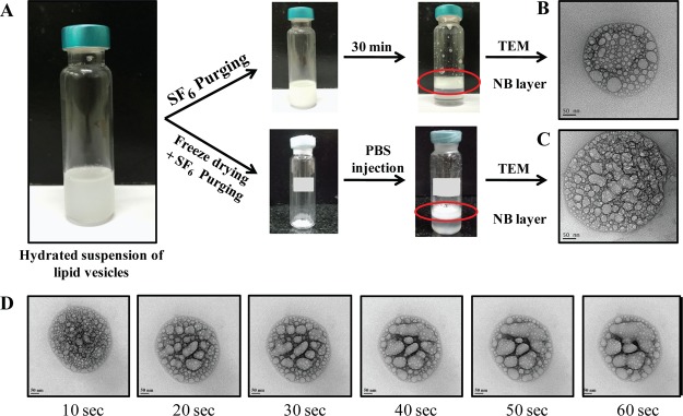
Figure 1.
[A] NB preparation showing the formation of compact middle layer for both freshly prepared and reconstituted NBs. Transmission electron micrographs of [B] freshly prepared NBs and [C] reconstituted NBs (scale bar—50 nm) showing the presence of spherical gas pockets. [D] Time lapse imaging of NBs under 200 kV electron beam showing coalescence of gas pockets.

