Figure 7.

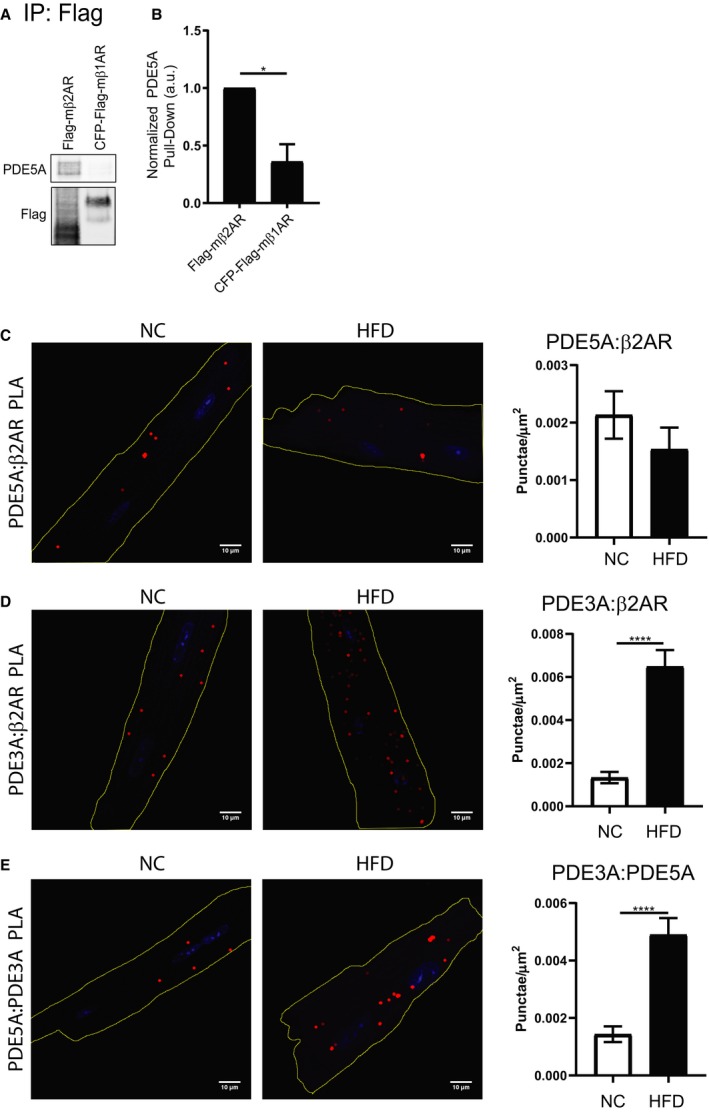
Phosphodiesterase 3 (PDE3) displays an increased association with the phosphodiesterase 5 (PDE5)–β2 adrenergic receptor (β2AR) protein complex in high fat diet (HFD) cardiomyocytes. A, Representative images and (B) quantification of coimmunoprecipitation of Flag‐tagged βARs from rabbit adult ventriculomyocytes (AVMs) expressing Flag‐mβ2AR or CFP (cyan fluorescent protein) – and Flag‐tagged mouse β1 adrenergic receptor (β1AR), with detection of PDE5A. n=3 rabbits. *P≤0.05 by unpaired t test. Proximity ligation assay (PLA) representative images and quantification in normal chow (NC) and HFD AVMs for the interaction between PDE5A and β2AR (C), between PDE3A and β2AR (D), and between PDE3A and PDE5A (E). Scale bar=10 μm. Red=PLA signal. Blue=DAPI. n=18 cells per group. ****P≤0.0001 by unpaired t test.
