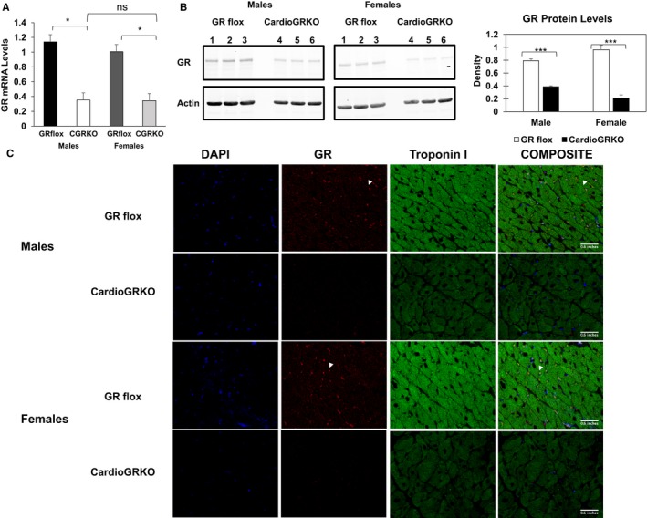
Figure 3.

GR mRNA and protein are significantly reduced in the hearts of cardiomyocyte‐specific glucocorticoid receptor knockout (cardioGRKO) mice and no glucocorticoid receptor (GR) is detected in the cardiomyocytes of the knockout mice. A, RT‐polymerase chain reaction of GR mRNA from hearts of control and cardioGRKO mice. B, Representative immunoblot show GR levels in control (GR flox) and knockout hearts. Hearts from male cardioGRKO have reduction of ≈70% in GR total protein levels, whereas female hearts present a reduction of ≈80% in GR protein levels. Immunoblots were quantified by densitometry using National Institutes of Health ImageJ analysis software. Data are mean±SEM (n=5 mice per group). *P<0.05; ***P<0.0001. C, Representative immunofluorescence staining of heart sections from control and cardioGRKO mice with anti‐troponin I (green) and anti‐GR (red) antibodies. DAPI is shown in blue. Data represent n=3 mice per sex and genotype. All pictures were acquired on a Leica TCS SP5 Spectral Confocal Microscope equipped with a ×40 (oil) objective. CardioGRKO indicates cardiomyocyte‐specific glucocorticoid receptor knockout; CGRKO, cardioGRKO; GR, glucocorticoid receptor.
