
Figure 2.
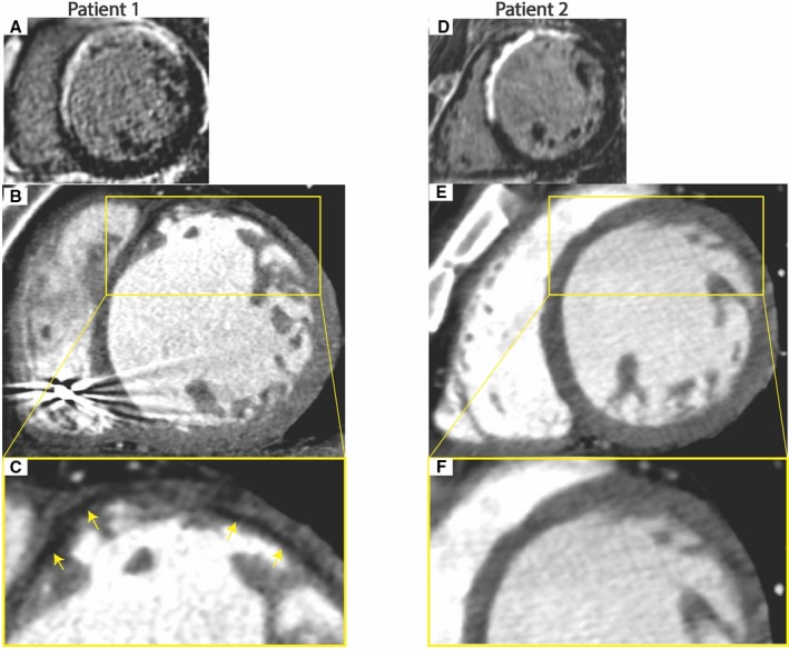
Examples of CT findings in patients with and without fat deposition with scar. A through C, Fifty‐eight‐year‐old man with VT 19 years after anterior infarction. Late gadolinium‐enhanced magnetic resonance imaging (MRI) shows antero‐septal scar (A). Arterial‐enhanced CT shows antero‐septal wall thinning with subendocardial fat deposition (yellow arrows) (B and C). D through F, 73‐year‐old man with VT 9 years after anterior infarction. Late gadolinium‐enhanced MRI shows antero‐septal scar (D). Arterial‐enhanced CT shows antero‐septal wall thinning with no fat deposition (E and F).