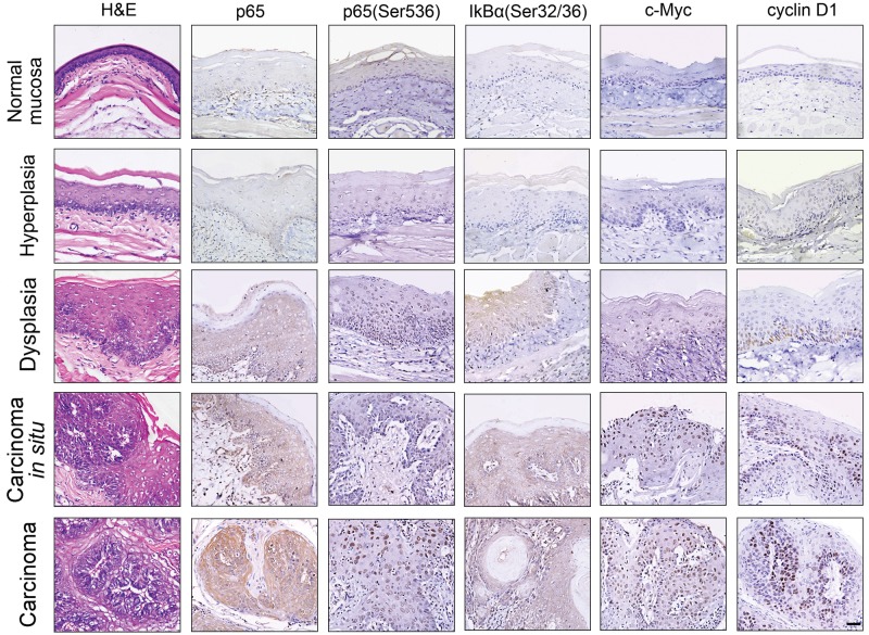
Figure 1.
NF-κB activation during chemically induced squamous cell carcinogenesis. The H&E staining and the immunohistochemical staining of total p65, phosphorylated p65 (Ser536), and IκBα (pSer32/36) or c-Myc and cyclin D1 at the stages of normal, hyperplasia, dysplasia, carcinoma in situ, and carcinoma in DMBA-induced squamous cell carcinogenesis of the hamster buccal pouch. Results are representatives of at least 3 tissue samples from more than 3 hamsters for each group. Scale bars, 50 μm.

