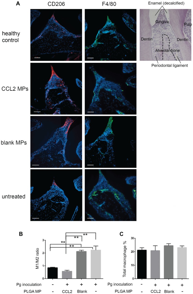
Figure 3.

The sustained CCL2 release formulation significantly upregulates the M2 phenotype macrophage ratio in gingival papilla in the Pg-induced mouse periodontitis model. (a) Representative immunofluorescence staining of M2 phenotype macrophages (CD206+; red) and all mature macrophages (F4/80+ green) in gingival papillae in sagittal sections of maxillary molars from BALB/c mice infected with Pg 60 d after inoculation. DAPI counterstain presents location of all cells. Scale bar = 50 um. Final magnification 200×. AB, alveolar bone; DP, dental pulp; GP, gingival papillae; M, molar. Quantitative analysis of M2 phenotype macrophage percentage in gingival papillae per high-power field and M1 phenotype macrophage percentage (b), as well as M1 phenotype:M2 phenotype ratio (c). After CCL2 MP treatment, M2 phenotype percentage was increased. M1 phenotype percentage and M1 phenotype:M2 phenotype ratio both decreased, while no changes were made on the total load of macrophages when compared with untreated control or blank MP control. n = 3; *P < 0.05 and **P < 0.005 by 1-way analysis of variance and a post hoc Tukey honestly significant difference test. Error bars indicate SD. CCL2, C-C motif chemokine ligand 2; M1, classically activated macrophage; M2, alternatively activated macrophage; MP, microparticle; Pg, Porphyromonas gingivalis.
