Figure 5.
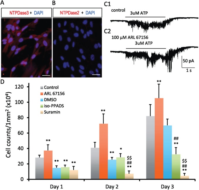
Expression of ectonucleotidases in hDPSCs and effects of purinergic signaling on hDPSC survival and proliferation. Immunofluorescence staining for ecto-NTPDases in hDPSCs. Expression of NTPDase3 was detected in hDPSCs (A). NTPDase2 was not detected in hDPSCs (B). Scale bars 20 μm. (C) Inhibition of ecto-NTPDases enhances purinergic signaling in hDPSCs. C1: ATP-induced (3 μM) inward current in hDPSCs. C2: Application of ecto-NTPDase inhibitor (100μM ARL 67156) enhanced the ATP-induced inward current in hDPSCs. D: Effects of purinergic signaling on the survival and proliferation of hDPSCs. DMSO exhibited deteriorating effects on survival and proliferation of hDPSCs. As compared with the control group, the cell number was significantly reduced at days 1, 2, and 3 in DMSO vehicle control group. Interestingly, inhibition of ectonucleotidase activity with 100mM ARL 67156 significantly increased the number of hDPSCs in culture from day 1 to day 3, which suggest trophic effects of ATP signaling on hDPSC survival and proliferation. As compared with the DMSO group (vehicle control), blocking P2X receptors with 100μM iso-PPADS had little effect on hDPSCs at days 1 and 2; an inhibition effect on hDPSCs was observed at day 3. Blocking P2X/P2Y receptors with suramin (100 μM) significantly reduced the number of hDPSCs in culture at days 1, 2, and 3 (cell proliferation). The data are presented as mean ± SD, n = 7 in each group (analysis of variance); least significant difference post hoc tests were performed at day 1, 2, or 3; and P < 0.05 was considered statistically significant. *P < 0.05, **P < 0.01, vs. control. ##P < 0.01, vs. DMSO. $$P < 0.01, vs. iso-PPADS. hDPSC, human dental pulp stem cell.

