
FIGURE 2:
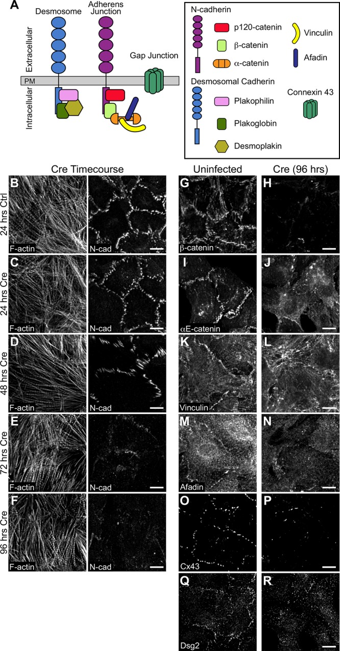
Loss of N-cadherin disrupts adhesion protein localization. (A) Cartoon schematic of desmosome, AJ, and gap junction proteins at cardiomyocyte cell–cell contacts. (B–F) Neonatal cardiomyocytes from Ncadfx/fx mice were uninfected (B) or infected with adenovirus expressing Cre recombinase (C–F) and fixed over 96 h to assess N-cadherin expression. Cells were stained for F-actin (left panel) and N-cadherin (N-cad, right panel). (G–R) Control and Cre-infected neonatal cardiomyocytes from Ncadfx/fx mice were fixed 96 h postinfection and stained for AJ proteins β-catenin and αE-catenin (G–J); AJ adapter proteins vinculin and afadin (K–N); connexin 43 (Cx43; O, P), and desmoglein 2 (Dsg2; Q, R). Images are maximum projections of 2–3 μm deconvolved stacks. Scale bar is 10 μm.