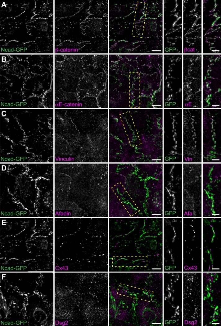
FIGURE 3:
N-cadherin-GFP rescues cardiomyocyte junctional complexes. (A–F) Neonatal Ncadfx/fx cardiomyocytes infected sequentially with adenoviruses expressing Cre and N-cadherin-GFP, fixed, and stained for AJ-associated proteins (A–D), gap junctions (E), and desmosomes (F). Individual and merged N-cadherin-GFP (green) and ICD components (magenta) channels are shown. Far right columns are higher magnifications of the boxed contact in the merge. Individual and merged channels are shown. Images are maximum projections of 2–3 μm deconvolved stacks. Scale bar is 10 μm in lower-magnification images, 5 μm in higher-magnification images.

